- Actinomycetes are a subclass of gram-positive bacteria that are distinguished by their capacity to produce spores and mycelia structures.
- Although they comprise an unique evolutionary line of creatures that includes everything from coccoid and pleomorphic forms to branching filaments, they exhibit noticeable chemical and morphological variety.
- These bacteria are highly significant to scientists, the pharmaceutical industry, and the agricultural sector since they have been shown to function as abundant reservoirs of therapeutic antibiotics.
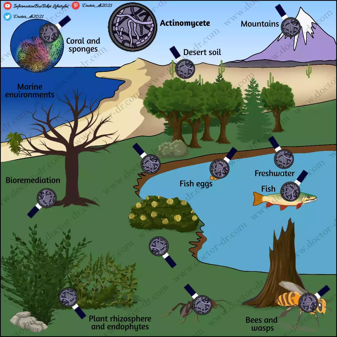
Actinomycetes that are free-living are common in soil habitats as well as in freshwater and marine ecosystems. They also have a significant ecological function in the recycling of organic matter. Many actinomycetes have developed symbiotic relationships with plants, fungi, animals, insects, and other organisms. The majority of these interactions between actinomycetes and their hosts are advantageous; actinomycetes either create NPs that enable their hosts to defend themselves against infections or pests or enzymes that break down tough natural polymers like lignocellulose.
Table of Contents
- Principle of Isolation of Actinomycetes
- Procedure of Isolation of Actinomycetes
- Expected Results of Isolation of Actinomycetes
Principle of Isolation of Actinomycetes
Actinomycetes is a non-taxonomic word for a class of common soil microorganisms sometimes known as "thread or ray bacteria." They are renowned for breaking down more durable organic compounds, such the complex sugar called chitin that is present in the exoskeletons of insects and other organisms. However, among other things, the type of soil, location, culture, and organic matter have a significant impact on the richness and variety of actinomycetes found in any given soil.
After dilution of the soil slurry, several agar medium can be employed to isolate Actinomycetes from soil sample. Isolation of Actinomycetes Agar is a commonly used selective medium. It has sodium caseinate as a source of nitrogen. In addition to being an amino acid, asparagus also contains nitrogen. In anaerobic fermentation, sodium propionate is employed as a substrate. The buffering mechanism is provided by dipotassium phosphate. The sulphates act as a source of metallic ions and sulphur. An other source of carbon is glycerol. By preventing mRNA translation, the fungi that are opportunistic are prevented from interfering further. Opportunistic fungus experience cell growth arrest and cell death as a result.
On the other hand, chitin, a complex sugar that is often hydrolyzed by Actinomycetes, is present in chitin agar. As a result, it can promote the growth of only actinomycetes while also suppressing the growth of other soil bacteria and fungi.
Procedure of Isolation of Actinomycetes
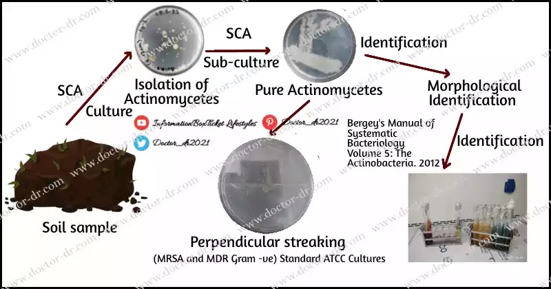
- To begin, make a soil slurry by combining 1 g of the collected dry soil with 10 ml of distilled water. For two minutes, a vortex is used to stir the slurry.
- The slurry is divided into duplicates and used to make four 1 in 10 fold serial dilutions.
- The agar plates (Actinomycete Isolation Agar supplemented with cyclohexamide (50 g/ml) and nystatin (50 g/ml) and Chitin agar/Starch Casein Agar) are then set up and 1 ml portions of each dilution are spread on them.
- Every spread plate is labelled before being incubated at 25 °C for 7 to 14 days.
- On day 14, colonies from both sets of cells are counted and noted in each dilution.
- To identify the diversity of the colonies that have developed, each plate is meticulously examined under a microscope. There are also calculations and records for the diversity, evenness, and richness indices.
- To generate pure single colonies, various colony types are then selected using sterile forceps and streaked out on glucose yeast extract agar plates.
- The streaked plates are then incubated, and the generation of antibiotics is evaluated afterwards.
Expected Results of Isolation of Actinomycetes
- Regarding Actinomycete Isolation. After 7 days of incubation on agar supplemented with cyclohexamide (50 g/ml) and nystatin (50 g/ml), actinomycete-specific pin-point white colonies can be seen with a distinct zone of inhibition around them.
- You can expect colonies to have a variety of appearances, including large bright white filaments with mycelia that resemble a net, pale white branching filaments that look powdery, dark brown uniformity and crumb-like appearance, light brown appearance with mycelia that resemble cilia on its boundaries, dark brown appearance with embedded concentric circular patterns, and yellow colonies with lovely mycelia and transparent boundaries.
- On chitin agar, certain actinomycete colonies have a distinct zone of hydrolysis surrounding them, making it easier to identify them macroscopically.


~1.webp)

.webp)
.webp)
