Table of Contents
- Structure of Polio Virus
- Genome of Polio Virus
- Epidemiology of Polio Virus
- Replication of Polio Virus
- Pathogenesis of Polio Virus
- Clinical Manifestations of Polio Virus
- Laboratory Diagnosis of Polio Virus
- Treatment of Polio Virus
- Prevention and Control of Polio Virus
Structure of Polio Virus
- The poliovirus belongs to the Picornaviridae virus family.
- Virions have a diameter of about 27 nm and are spherical in form.
- The particles are made up of a protein shell encircling a naked RNA genome, making them simple particles.
- The genome is monopartite, linear, ssRNA(+), polyadenylated, and contains a single ORF that codes for a polyprotein. Its size ranges from 7.2 to 8.5 kb.
- The four basic proteins VP1, VP2, VP3, and VP4 make up the capsids.
- The protomer, which has one duplicate of each of the three capsid proteins (VP1, VP2, and VP4), is the fundamental component of the picornavirus capsid.
- VP1 to VP3 combine to create the shell, and VP4 is located on the interior surface.
- The viral particles have no lipid envelope, and organic solvents have no effect on their capacity to spread infection.
Genome of Polio Virus
The DNA of the poliovirus is composed of three parts:
- Uncapped, making up about 10% of the genome, the 5′ noncoding region (NCR) is covalently attached to the viral protein VPg at its terminus.
- A single open reading frame, dubbed P1 for capsid proteins and P2 and P3 for nonstructural proteins, appears to encode all of the viral proteins.
- A 3′ NCR that ends in a polyA tip is brief.
- The length of the chromosomes ranges from 7,209 to 8,450 bases.
- Internal ribosome entry site (IRES), a component that controls mRNA translation by internal ribosome binding, is present in the 5′-noncoding region.
- The regions P1 comprise the 1A-VP4, 1B-VP2, 1C-VP3, and 1D-VP1 segments for the structural proteins that make up the capsid protein.
- Three non-structural proteins, 2A, 2B, and 2C, make up P2 and are involved in viral reproduction.
- Four non-structural proteins make up P3.
- The reproduction complex is anchored to the cell membrane by 3A.
- It is VPg protein, 3B.
- Cysteine protease is the enzyme that separates the protein from the polypeptides in 3C.
- RNA reliant RNA polymerase is used in 3D.
Epidemiology of Polio Virus
- Three epidemiologic stages have been observed for poliomyelitis: endemic, epidemic, and vaccine era.
- Poliomyelitis was present everywhere prior to the start of global eradication attempts; it was year-round in tropical regions and seasonal in temperate regions.
- A winter epidemic was unusual.
- The illness affects people of all ages, but because adults have developed acquired immunity, toddlers are typically more vulnerable than adults.
- Poliomyelitis, also known as "infantile paralysis," affects infants and young children in poor countries where the environment is conducive to the widespread spread of the virus.
- Prior to vaccination, the age distribution changed in developed nations, with most patients being older than 5 years old and 25% being older than 15 years.
- The case fatality rate varies and can range from 5% to 10%. It is greatest in the oldest patients.
- The number of paralytic poliomyelitis cases per year was around 21,000 prior to the start of vaccination programmes in the US.
- The only known source of transmission is humans.
- In temperate regions with high levels of hygiene, epidemics have been followed by lulls in viral spread until enough susceptible young people have reached adulthood to create a reservoir for transmission in the area.
Replication of Polio Virus
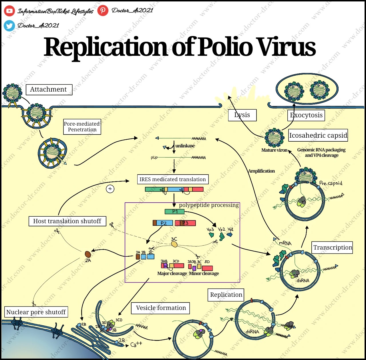
- The virus uncoils its DNA after binding to a cellular receptor.
- The virus RNA is depleted of VPg before being translated.
- Individual virus proteins are produced through nascent cleavage of the polyprotein.
- Membrane compartments are where RNA is created.
- The viral RNA polymerase copies viral (+) strand RNA to create full-length (-) strand RNAs, which are then copied to create more (+) strand RNAs.
- Translation of freshly synthesised (+) strand RNA early in infection results in the production of extra viral proteins.
- The (+) strands join the morphogenetic pathway later in the infection.
- Viral particles that have just been created are expelled from the cell by lysis.
Pathogenesis of Polio Virus
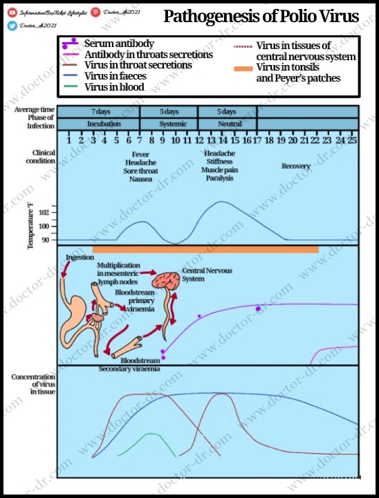
- When contaminated water is consumed, the virus enters the body through the mouth via the faecal oral pathway.
- In the beginning, the oropharynx and gastrointestinal mucosa of the virus proliferate.
- Before symptoms appear, the virus is frequently found in the pharynx and stools.
- The protease, other digestive enzymes, and bile lytic activities cannot harm viruses because they are immune to stomach acidity.
- The tonsils and Peyer's patch of the ileum are the first parts of the body the virus enters and spreads to.
- A 9–12 day incubation time is required.
- Following this, the virus spreads to nearby lymph nodes before entering the blood and producing primary viremia.
- Early in the illness, typically before paralysis sets in, antiviral antibodies start to manifest.
- To stop an infection from expanding, antibodies are created.
- Invading the bloodstream and producing secondary viremia, the virus multiplies and infects the ReticuloEndothelial System (RES) over time.
- The poliovirus crosses the blood–brain barrier and enters the brain during this time of viremia.
- The virus particularly combines with neural cells, demonstrating tissue tropism.
- The dorsal root ganglia, motor neurons, and anterior horn of the spinal cord all have receptors that the virus can detect.
- Paralysis results when motor nerves are lost.
- Virus infection of the brain stem results in bulbar poliomyelitis.
Clinical Manifestations of Polio Virus
- The initial symptoms of viremia, which last for one to five days, include temperature, malaise, headache, drowsiness, constipation, and sore tongue.
- The incubation phase lasts for 10 days on average, but it can also be 4 days or 4 weeks.
1. Asymptomatic illness
- It results from a viral illness that is limited to the intestine and oropharynx.
2. Abortive poliomyelitis
- It is a minor disease that affects about 5% of those who are infected.
- It is a febrile illness that includes vomiting, nausea, headaches, sore throats, lack of appetite, and abdominal discomfort.
- Typically, neurological signs are nonexistent.
3. Non paralytic poliomyelitis
- Some individuals who experience polio symptoms get a type of polio that doesn't cause paralysis (abortive polio).
- The mild, flu-like signs and symptoms that are characteristic of other viral illnesses are typically caused by this.
- Fever, sore throat, headache, vomiting, fatigue, back pain or stiffness, neck pain or stiffness, pain or stiffness in the limbs or legs, and muscular weakness or tenderness are just a few of the signs and symptoms that can last up to 10 days.
4. Paralytic poliomyelitis
- Initial paralytic polio symptoms, such as temperature and headache, frequently resemble non-paralytic polio symptoms.
- But after a week, additional symptoms start to show, such as: Reflex loss, excruciating muscular pain or weakness, and shaky, droopy limbs (flaccid paralysis)
5. Post poliomyelitis syndrome
- Some individuals experience post-polio syndrome, a collection of incapacitating signs and symptoms, years after they had polio.
- Common indications and symptoms include progressive joint pain and weakness, exhaustion, muscular atrophy, difficulty breathing or swallowing, breathing disorders associated with sleep, such as sleep apnea, and a decreased tolerance for cold temps.
6. Bulbar poliomyelitis
- The participation of the cranial nerves, most frequently the 9th, 10th, and 12th, is what causes this.
- The pharynx muscles, vocal cords, and respiration are often affected, making this disease more serious.
- 75% of patients who have the disease run the risk of dying.
Laboratory Diagnosis of Polio Virus
Specimen: stool, rectal swab, throat swab, CSF (rare)
1. Microscopy
- By using immune electron microscopy or direct electron microscopy, viruses can be found in faeces samples.
- Although viral is infrequently seen in CSF, lymphocytic pleocytosis is primarily seen when CSF is examined under a microscope.
2. Virus isolation
- Feces and pharyangeal desires may yield the virus.
- By cultivating on monkey kidney, human amnion, HeLa, Hep-2, Buffalo green monkey (BGM), MRC-5, and other cell cultures, viruses are isolated from excrement and throat swabs.
- In 3-6 days, cytopathogenic consequences become apparent.
- Cell retraction, increased refractivity, cytoplasmic granularity, and nuclear pyknosis are a few examples of cytotopathic impacts.
- By neutralising an isolated virus with a particular antiserum, it can be recognised and typed.
3. Serodiagnosis
- Evidence of a fourfold rise in antibody titer in serum samples taken during an acute illness and during a period of recovery.
- To prove the existence of antibodies, neutralisation and complement fixation tests are conducted.
4. Molecular diagnosis
- Virus detection can also be accelerated using polymerase chain reaction (PCR) tests.
Treatment of Polio Virus
- There are no antiviral medications available to cure poliomyelitis.
Prevention and Control of Polio Virus
- To lower the risk of transmission in endemic countries, better sanitation, hygienic practises, and water availability are crucial.
- The mainstay of the fight to eradicate polio is immunisation, and both live and killed viral vaccines are available.
- Salk, a vaccine made from viruses produced in monkey kidney cultures, is formalin-inactivated.
- The vaccine for a killed virus elicits humoral antibodies but not local intestinal immunity, allowing the virus to continue to grow in the gut.
- In main monkey or human diploid cell cultures, the live attenuated vaccine (Sabin) is grown before being administered orally.
- The live polio vaccine grows within the host, immunises it against virulent strains, and spreads infection.
- The vaccine results in the production of secretory IgA antibodies in the intestine in addition to immunoglobulin M (IgM) and IgG antibodies in the blood, allowing mucosal immunity.
- Live and killed viral vaccines both produce antibodies and shield the CNS from future wild virus invasion.
- In the past, oral polio vaccine was the most widely used vaccine in global campaigns, and it is still used in endemic regions.
- It benefits from inducing intestinal and humoral immunity as well as being affordable and simple to give.
- However, after receiving the live-virus vaccine, the gut exhibits a much higher level of resistance, suggesting that it may serve as a potential interference limiter for oral vaccines.
- The drawback is the negligible chance of vaccine-associated paralytic poliomyelitis (VAPP), which affects 4 out of every 100,000 immunised children and unvaccinated contacts.
- The intramuscular injection of inactivated poliovirus vaccine carries no danger of VAPP.
- Inactivated vaccine has the drawback of not granting intestinal immunity, being ineffective at containing outbreaks, being more costly, and requiring better-trained personnel for delivery.
- Over the past few decades, OPV use in European nations has progressively given way to IPV use, and all EU Member States now use IPV in their childhood immunization programmes.


%20Technique%20A%20Comprehensive%20Guide%20to%20Water%20Quality%20Testing%20and%20Microbiological%20Analysis.webp)
~1.webp)

.webp)
.webp)

