Table of Contents
- What is Sporothrix schenckii?
- Habitat of Sporothrix schenckii
- Morphology of Sporothrix schenckii
- Cultural Characteristics of Sporothrix schenckii
- Transmission of Sporothrix schenckii
- Pathogenesis of Sporothrix schenckii
- Virulence Factors of Sporothrix schenckii
- Clinical Manifestations of Sporotrichosis
- Diagnosis of Sporotrichosis
- Treatment of Sporotrichosis
- Prevention and Control of Sporotrichosis
What is Sporothrix schenckii?
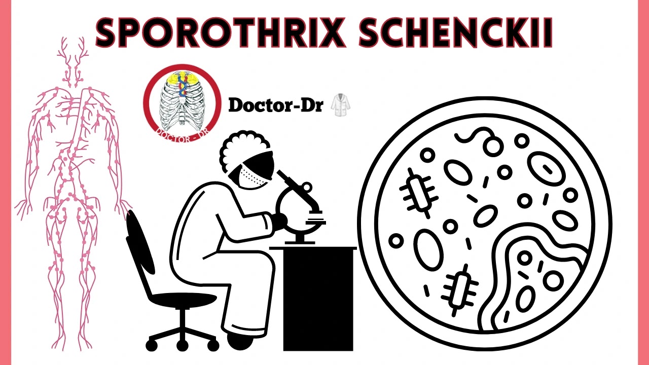
A common dimorphic fungus found in soil, on living things, and in decaying habitats is Sporothrix schenckii.
- It is a pathogenic fungus that affects both people and animals and produces sporotrichosis.
- A persistent mycotic infection that affects the cutaneous and subcutaneous tissues is sporotrichosis.
- The development of tiny, ulcerative, and suppurative lesions on and in the skin tissues is linked to the fungal infection.
- Additionally, nodular lesions that can develop into ulcerative and suppurative lesions might have an impact on the lymphatic system.
- Through skin abrasions and occasionally through inhalation into the lungs, the fungus enters the host.
- Rarely are there signs of subsequent spread to the muscles and bones, while there have been isolated cases of infection of the lungs, genitourinary tract, and central nervous system.
- Benjamin Schenck, a medical student at the Johns Hopkins Hospital in Baltimore, Maryland, isolated Sporothrix schenckii for the first time in 1896 from a 36-year-old male patient who had lesions on his right hand and arm.
Habitat of Sporothrix schenckii
- Soil is a frequent habitat for this fungus.
- Additionally, it may be discovered on living plants like roses and barberry bushes as well as in plant remains like mulch made of pine bark and sphagnum moss.
- At low temperatures and in the environment, Sporothrix schenckii develops as a mould.
- In living organisms and at temperatures over 35 °C, it changes into a yeast form.
- For florists, gardeners, and forestry workers, sporotrichosis—an infection brought on by Sporotrix schenckii—is a work-related illness.
- Tropical and subtropical regions frequently have it.
Morphology of Sporothrix schenckii
- A dimorphic fungus is called Sporothrix schenckii.
- It exhibits a filamentous shape in the fundamental mycological culture at 25°C made up of hyaline, septate hyphae that are 1 to 2 m broad.
- The colonies' fungal development is characterised by branching septate hyphae that create tiny, distinct, brown conidia that measure between three and five microns in diameter.
- Conidiophores, which emerge at right angles from the septate hyphae, are responsible for producing the conidia.
- The tips of the conidiophore are tapered.
- At the apex of the conidiophore, where the produced conidia are grouped on small denticles, a flower-like look is created.
- The conidia are smooth-walled, hyaline, single-celled, ovoid or elongated, measuring 3-6 x 2-3 m.
- As the fungal culture ages, large, singularly occurring conidia may also be generated.
- The big conidia are obovate to angular, with thick, black walls.
- In rich medium, spherical or oval-shaped fusiform budding yeast cells with a size of around 1-3 3-10 m are generated.
Cultural Characteristics of Sporothrix schenckii
- At 25°C, they slowly develop in common mycological agar medium like malt extract agar and potato dextrose agar, generating lustrous, blackish/greyish colonies that eventually mature into moist, glabrous, wrinkled, and fuzzy colonies that are coloured black.
- Sporotrix schenckii is thermally dimorphic in a rich medium like brain heart infusion media (BHI) at 35-37°C, and its development is characterised by many budding yeast cells.
- They create colonies of glabrous yeast cells in the BHI medium that range in colour from white to yellow.
Transmission of Sporothrix schenckii
- By traumatically implanting the fungus from contaminated soil, plants, and organic materials, the fungus gets inoculated into the host.
- Florists, gardeners, miners, and forest workers frequently experience it.
- A few cases in Brazil have also been documented of zoonotic transmission through cats.
- It takes 1 to 12 weeks for it to incubate.
Pathogenesis of Sporothrix schenckii
Virulence Factors of Sporothrix schenckii
Thermotolerance
- Growing conditions for Sporothrix schenckii range from 35 to 37 °C.
- At 35°C, it can result in lymphatic sporotrichosis, and at 37°C, it can result in extracutaneous and widespread sporotrichosis lesions.
Melanin synthesis
- The capacity to produce melanin, an insoluble substance that has been connected to the pathogenicity of several fungal families, is possessed by Sporothrix schenckii.
- The dematiaceous conidia of the fungus contain melanin.
- Sporotrix schenckii's resistance to macrophage phagocytosis is increased by conidial melanization, which also triggers the initial stage of infection by the conidia spores, the infectious particle of the fungus.
- Because it makes the fungus more invasive into the host, melanin pigmentation plays a significant role in the development of cutaneous sporotrichosis.
Adherence
- Sporotrix schenckii is able to recognise the glycoproteins on the extracellular matrix of the cutaneous tissues, such as fibronectin, laminin, and type II collagen, thanks to adhesins such integrins and adhesin lectin-like molecules.
- The yeast cells' surfaces include fibronectin adhesins, which are responsible for the host-fungal adhesion factors.
- On yeasts and fungal hyphae, laminin receptors may be found that can bind to the extracellular matrix.
- These adhesins facilitate fungal adhesion to host tissues and promote the spread of illness.
Clinical Manifestations of Sporotrichosis
Lymphocutaneous infection
- It is Sporotrix schenckii's most frequent symptom.
- The hands and arms, lower limbs, trunk, and face all have lesions.
- Within the first several weeks after a fungal inoculation, primary lesions appear.
- Small nodules that develop into ulcers are the lesions.
- The lesions are non-pruritic and somewhat uncomfortable.
- The lymphatics are affected by new nodules that spread and ulcerate.
- The lymph nodes may swell and hurt if lymphangitis develops.
Cutaneous Infection
- The suppurative and granulomatous inflammatory reaction in the dermis and subcutaneous tissue is frequently caused by Sporothrix schenckii.
- Microabscess, fibrosis, hyperkeratosis, parakeratosis, and pseudoepitheliomatous hyperplasia are its defining features.
Fixed Cutaneous Infection
- This is the growth of a single lesion that would typically appear on the face.
- The lesion might be verrucous or ulcerated.
- Only antifungal medication can make the lesion go away.
Osteoarticular sporotrichosis
- Sporotrichosis is an uncommon illness that often affects drinkers.
- Septic arthritis, which develops as a result of traumatic inoculation and spreads to the joints, is what distinguishes it.
- Carpal tunnel syndrome can be accompanied with bursitis and tenosynovitis.
Pulmonary sporotrichosis
- It is an infection, either subacute or chronic.
- People with Chronic Obstructive Pulmonary Disease (COPD) frequently experience it.
- After conidia are inhaled, it happens.
- Fever, exhaustion, loss of weight, coughing, sputum production, and hemoptysis are its hallmark symptoms.
- Infected people frequently have TB, atypical mycobacterial infection, or chronic cavitary pulmonary histoplasmosis.
Diagnosis of Sporotrichosis
Specimen: Tissue biopsy, pus from lesions, sputum, urine, blood, and cerebrospinal and synovial fluids.
Direct Examination
- 10% potassium hydroxide (KOH) wet mount to observe for budding yeast cells
- Gram staining stains the yeast cells gran positive.
- Sporothrix schenckii conidiophores and conidia
Histological Examination
- Hematoxylin and eosin (H&E) stain,
- Gomori methenamine silver (GMS)
- Periodic acid-Schiff (PAS) stain
Cultural Examination
- Growth happens in 5-7 days at 25°C on saboraud agar with chloramphenicol and on medium with cycloheximide, such mycobiotic agar, creating filamentous hyaline colonies that get black at the centre as the culture ages. Additionally, dematiaceous conidia are seen.
- To demonstrate conidiogenesis, potato dextrose agar and cornmeal agar are utilised.
- Heart and Brain Infusion At 35 to 37°C, agar, chocolate agar, and blood agar are utilised to demonstrate dimorphism. Within 5-7 days, colonies grow to generate yeast cell colonies that range in colour from creamy and yellow to tan.
Molecular Detection
- Detection of PCR amplicons
- Oligonucleotide primers for differentiation of Sporortrix schenckii from other fungal species.
Serological Test
A high titer agglutinin antibody test is performed in a test tube.
Antigen-coated latex particles are used in latex agglutination to find sporotrichin antigen in patients' serum
Sporotrichin Skin Test
- This examination is designed to find cellular immune responses with delayed hypersensitivity.
- It has the ability to identify and confirm both recent and old Sporothrix schenckii infections.
- Bulbar conjunctival sporotrichosis has been diagnosed using the sporotrichin skin test.
Treatment of Sporotrichosis
- Oral administration of a saturated solution of potassium iodide
- Itraconazole is currently the first-choice treatment
- Systemic infection can be treated with amphotericin B
Prevention and Control of Sporotrichosis
- putting on safety gear like gloves and long sleeves when performing high-risk tasks like handling sphagnum moss, wires, rosebushes, hay bales, conifer (pine) seedlings, or other items that might promote the exposure to the fungus
- To stop the spread of zoonotic diseases, sporotrichosis in cats must be properly treated and housed in isolation.


~1.webp)

.webp)


