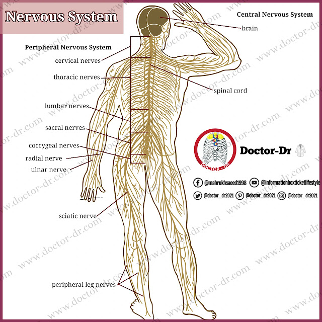
Your nervous system is a multi-organ, multi-nerve system that manages all of your thoughts and actions. Figure 13 displays a thorough diagram of the human nervous system. The brain is the control center of the neurological system. Nerve cells in the brain receive messages from every region of the body. The brain analyses these messages and sends a reaction back to other regions of the body through additional nerve cells. The spinal cord is where most of the messages are transmitted.
You can react to environmental changes thanks to your nervous system. For use days, months, or even years from now, your nervous system stores information. The voluntary and involuntary muscle movements of your body are also controlled by it. Your muscles will tense and relax as a result. Because there are billions of nerve cells in your body, your nervous system is able to do all of these tasks. Nerve cells communicate with one another and with muscles using signals.
Figure 13 The Nervous System
Nerve Cells
The basic unit of the nervous system is the neuron. It is frequently called a nerve cell. Neurons are the messenger cells that send information throughout your body. Impulses are the name for these sensory and informational transmissions. Neurons make up the information storage and control system for the body's numerous tasks. Although there are many types of neurons, there are certain properties that are identical in all types.
Structure of Nerves
Figure 13-1 depicts the cell body of a neuron and the fibres that go to and from it. One axon and one or more dendrites make up each nerve fibre.
The axon is a long fibre that sends impulses to other neurons outside of the cell body. Axons that connect distant neurons in your body may extend more than one yard. A bundle of axons from several neurons make up a nerve. More than 1000 axons may be connected in one neuron. Acting independently of the others, each axon.
The dendrites are fibers like tiny trees that receive impulses and send them to the cell body. They are too small to be seen without a microscope. Because dendrites have many branches. one neuron can receive messages from hundreds of other neurons.
Figure 13-1 Neuron structure
Nerve Impulses
The impulse that travels along a neuron is similar to a tiny electrical charge. Along an axon, nerve impulses can move as quickly as 360 feet per second. An impulse travelling at that speed would take under a second to complete a football field's length.
A synapse [SIN aps] is a gap that exists between the dendrite of one neuron and the terminal of its axon. Neurotransmitters convey an impulse from the terminal of an axon to the dendrite of the following neuron. Chemicals called neurotransmitters (nur oh TRANS mitur) go from an axon to a dendrite across a synaptic gap. An impulse is started by neurotransmitters, which then travels up the axon and down the dendrite to the cell body.
Kinds of Neurons
Neurons come in three different subtypes. The relationships between the various types of neurons are shown in Figure 13-1. Sensory neurons are the brain's internal and external environment change detectors. They transport nerve signals to the spinal cord or brain from the internal organs, muscles, skin, and sensory organs. For instance, the neurons in your body that detect cold transmit this signal to your brain when you touch an ice cube.
Interneuron neurons are those that receive sensory information and respond. Only the brain and spinal cord contain them. In the aforementioned example, several interneurons in your brain work in concert to help you become aware that you are touching an ice cube. The motor neurons receive the information, which causes the muscles to move.
The body's muscles, glands, and internal organs get signals from the interneurons via the motor neurons. Motor neuron signals are sent to the muscles and regulate every movement of the body, from a blink of the eyelids to a leap into the air. In the case of the ice cube, the motor neurons' message may instruct the hand's muscles to contract in order to pick up the ice cube and put it in a glass.
Key points:
- Neurons are the basic unit of the nervous system, also called nerve cells.
- Neurons transmit information through impulses.
- A neuron consists of a cell body, dendrites, and an axon.
- Axons send impulses to other neurons outside the cell body, while dendrites receive impulses and send them to the cell body.
- Nerve impulses move as fast as 360 feet per second along an axon.
- Synapses are the gaps between the dendrite of one neuron and the terminal of its axon.
- Neurotransmitters are chemicals that convey impulses from the terminal of an axon to the dendrite of the following neuron.
- There are three types of neurons: sensory neurons, interneurons, and motor neurons.
- Sensory neurons detect changes in the internal and external environment and transmit nerve signals to the spinal cord or brain.
- Interneurons receive sensory information and respond, located only in the brain and spinal cord.
- Motor neurons regulate movements of the body's muscles, glands, and internal organs.
The Central Nervous System
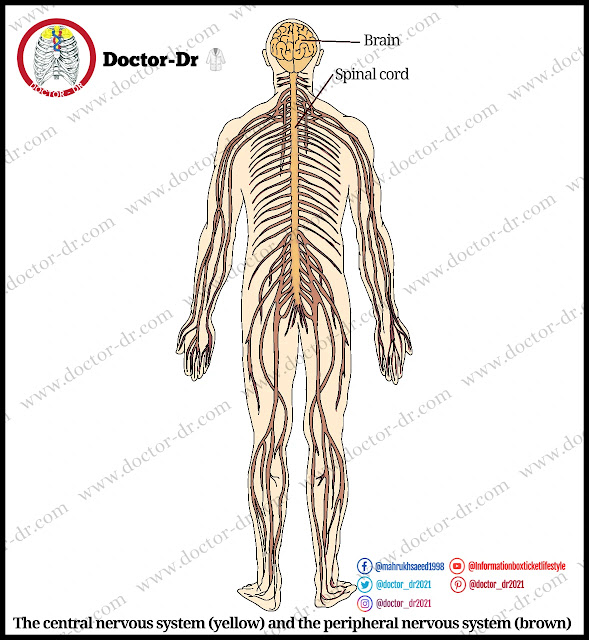
In the past, people believed that the heart managed the body. The brain is now understood to be the body's primary control system. It is interesting but yet poorly understood how this three pound mass of spongy tissue is able to receive, retain, and convey information. The central nervous system is made up of the brain and spinal cord. Each component of the brain and spinal cord plays a part in the central nervous system's operations.
Protective Coverings
The brain and spinal cord have multiple protective coverings because they are so important. Bone makes up the outside layer. The brain is encased in and shielded by the skull. The spinal cord is encased in the vertebrae. Three membranes under the skull and vertebrae offer further protection. In addition, a material known as cerebrospinal fluid (ser wh broh SPY null) fills some areas in the brain as well as the spaces between the middle and inner membranes. In the case of a hit or an abrupt change in direction, this fluid protects the brain and spinal cord.
Figure 13-2 The central nervous system (yellow) and the periph- eral nervous system (brown)
The Cerebrum
Figure 13-3 Control centers at the brain.
The cerebrum (cerebrum) is the biggest and highest region of the brain, which controls your ideas and behaviour. The highly developed intelligence of humans is a function of the cerebrum. Figure 13-3 illustrates how the cerebrum's surface resembles a ruffled walnut with many grooves. In healthy brains, these grooves follow a pattern. The patterns are used to pinpoint certain brain areas. Some areas get information about your movements or what you can smell, hear, and see. Your capacity to think, write, speak, and express emotions is governed by other portions. A breakdown of the brain's many areas is shown in Figure 13-3.
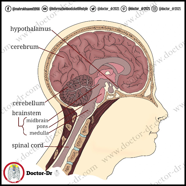
Figure 13-4 The structure of the brain.
The cerebrum's outer layer is known as the cerebral cortex (suh REE brull). It is made up of interneuron cell bodies. About three-fourths of the nerve cell bodies in your nervous system are located here. Because the nerve cell bodies of the cerebral cortex are grey, it is referred to as grey matter. The part of the brain below the cortex that is referred to as "white matter" is called that. The white matter contains billions of axons that branch off from and link to the cortex. In the cerebrum's deepest part, the thalamus and hypothalamus are situated. The thalamus is the region of the brain that communicates sensory information to the cerebral cortex.
The region of the brain known as the hypothalamus controls a number of the body's most fundamental functions, including body temperature, sleep, digestion, and hormone secretion. Figure 13-4 depicts these structures. The front and rear halves of the cerebrum are split in half. The opposing sides of the body are controlled by the two halves of the cerebrum. The muscles on the left side of the body are truly under the direction of the brain's night side. The muscles are managed by the left side of the brain.
The Cerebellum
The cerebellum (sehr uh BEL um) is the area of the brain that controls the muscles you employ to perform actions like running and walking. A steady stream of nerve impulses from the body and the cerebrum are coordinated by the cerebellum, which is located under and below the cerebrum. In order for your muscles to respond the way you want them to, the cerebellum combines this information. Additionally, it aids in posture maintenance and balance management.
The Brainstem
The brainstem is the part of the body that links the spinal cord to the brain. Figure 13-4 depicts the three divisions of this stem. The medulla, pons, and midbrain are located at the summit of the stem. Just above the spinal cord is where the medulla is located. The centre that regulates some of life's most crucial processes, including breathing, blood pressure, heart rate, and swallowing, is known as the medulla (muh DUL uh). You breathe more forcefully and your heart beats more quickly when you're anxious or enthusiastic. The medulla is stimulated by your senses and your brain's emotional regions.
The cerebellum and the cerebrum, as well as the right and left sides of the brain, are connected by a bundle of nerve fibres called the pons. The pons aids in respiratory regulation and eye movement control. A collection of nerves in the midbrain regulates several involuntary behaviours, including pupil size and eye movements.
The Spinal Cord
Within the vertebral column is the main nerve trunk of the body, the spinal cord. It reaches immediately below the ribs from the medulla. All of the nerves in your body are connected to the central nervous system through nerve fibres in the spinal cord, which extend to every area of the body. The body may not be able to move or feel from the site of injury downward if the spinal cord is destroyed. A cross section of the spinal cord and the placement of its protective coverings are shown in Figure 13-5. The grey matter is located inside the spinal cord, as you can see.
Figure 13-5 Structure of Spinal Cord
Key points:
- The brain is the body's primary control system, and the central nervous system is made up of the brain and spinal cord.
- The brain and spinal cord have multiple protective coverings, including bone, membranes, and cerebrospinal fluid.
- The cerebrum is the biggest and highest region of the brain, controlling ideas and behavior, and is made up of the cerebral cortex (grey matter) and white matter.
- The thalamus communicates sensory information to the cerebral cortex, and the hypothalamus controls fundamental bodily functions.
- The cerebellum coordinates nerve impulses from the body and cerebrum to control muscles and balance.
- The brainstem links the spinal cord to the brain and is composed of the medulla, pons, and midbrain, controlling vital processes such as breathing, blood pressure, and eye movements.
- The spinal cord is the main nerve trunk of the body, extending to every area of the body and connecting all nerves to the central nervous system.
The Peripheral Nervous System
The peripheral nervous system is the system that transmits signals from the body to the brain. The two nerve systems are in continual communication with one another. The spinal cord's 31 pairs of big nerve branches and the brain's 12 pairs of nerve branches allow for this. Numerous thousands of sensory and motor nerve fibres are present in these nerves, which connect to every area of the body. Information from the senses is transported through the peripheral nervous system. This includes awareness-based experiences like touch. Additionally, it transmits feelings that you are not aware of, such pressure on your tendons. Motor impulses that regulate voluntary activity are carried by a portion of the peripheral nervous system.
The autonomic nervous system [aw tuh NAHM ik] is the portion of the peripheral nervous system that regulates involuntary reactions. For instance, automatic reactions regulate your digestion and respiration. The majority of the motor nerves that make up the autonomic nervous system.
Numerous organs are impacted by the autonomic nervous system. These include the kidneys, liver, stomach, intestines, sweat glands, salivary glands, and eye pupils. There are two sections to the autonomic nervous system. The two components of the autonomic nervous system have different effects on the identical organs that they impact. The autonomic nervous system's parasympathetic division is responsible for typically slowing down bodily processes. As an illustration, while you are sleeping. The heart rate and breathing rate are slowed by it. When you are sleeping, it is most active.
The portion of the autonomic nervous system that is active or under emotional stress is known as the sympathetic nervous system. You're probably relaxed and motionless while you read this. Your heart rate is being controlled by parasympathetic impulses, keeping it slow and constant. Your sympathetic nervous system would play a bigger role if something excited you or made you move swiftly. Your breathing would become more rapid and your pulse rate would quicken. Blood flow in organs including the stomach and intestines would be reduced. Blood supply to working muscles would also rise at the same time. The two components of the autonomic nervous system interact to maintain your body functioning normally at all times. Without your conscious involvement, it happens. The interaction of the sympathetic and parasympathetic nerve systems is shown in Figure 13-6.
Figure 13-6 Action of the autonomic nervous system.
Key points:
- The peripheral nervous system transmits signals from the body to the brain.
- It consists of 31 pairs of nerve branches in the spinal cord and 12 pairs in the brain.
- It carries sensory information (touch, pressure, etc.) and motor impulses (voluntary activity).
- The autonomic nervous system regulates involuntary reactions, such as digestion and respiration.
- It impacts organs such as the kidneys, liver, stomach, intestines, sweat glands, salivary glands, and eye pupils.
- The autonomic nervous system has two components: the parasympathetic division and the sympathetic division.
- The parasympathetic division slows down bodily processes and is most active during sleep.
- The sympathetic division is active under emotional stress and prepares the body for action.
- The two components of the autonomic nervous system interact to maintain normal body function.
Lesson Review
Your nervous system is a complicated collection of muscles, glands, and nerves that manages all of your bodily functions. All of the body's parts get impulses thanks to the cooperation of the nervous system's two major components. Your muscles, balance, posture, and other crucial bodily processes are all coordinated by the brain, which also controls your thoughts.
- What is a synapse?
- Name three kinds of neurons.
- Define gray matter.
- Where would you find the medulla, pons, and midbrain?
- 5 Which part of the autonomic nervous system has a stronger role in responding to emotional stress?


~1.webp)

.webp)
.webp)
