Table of Contents
- Flagella
- Structure of Flagellum
- Flagella formation mechanism
- Types of Flagella
- Bacterial flagella arrangement
- Functions of Flagella
- Examples of Flagella
Flagella
A flagellum, or flagella, is a slender, lash-like structure found on the cell body, playing crucial roles in various cellular functions.
- The term "flagellum" originates from the Latin word for whip, underscoring its long, slender resemblance to this tool.
- While flagella are distinctive in Mastigophora protozoans, they are also present in diverse microscopic and macroscopic organisms such as bacteria, fungi, algae, and animals.
- Primarily serving as a locomotion organelle, flagella are vital for movement and contribute to activities like food gathering and circulation in different organisms.
- Flagella exhibit variations in structure, mechanism, movement, and functions across different living entities. In archaea, for instance, the term "archaellum" is used to describe a distinct flagellum that differs from bacterial flagella.
- Although flagella share structural similarities with cilia, they differ in terms of number, occurrence, movement, and occasionally, functions.
- Researchers have extensively explored these appendages in various animal groups, revealing their roles in both movement and sensation. Flagella can detect changes in environmental composition and pH.
- Among the well-studied flagella, the one found in Chlamydomonas reinhardtii is particularly noteworthy due to its size.
- While flagella commonly appear at the polar ends of cells, their number and positioning vary, maintaining consistent composition and functions within a species.
Structure of Flagellum
The size, structure, and quantity of flagella vary between prokaryotes and eukaryotes, with notable distinctions even within prokaryotes, such as the differences between bacterial and archaeal flagella. Similarly, the composition and process of flagella formation exhibit diversity. Nevertheless, the fundamental structure of a flagellum shares common elements across all domains of life.
The fundamental components of a flagellum encompass:
1. Filament:
- The filament, constituting approximately 98% of the flagellum's structures, is its most conspicuous part.
- Emerging from a hook-like structure within the cell's cytoplasm, the filament has an average length of 18 nm.
- Filament lengths vary among different organisms; for instance, bacterial flagella filaments measure 20 nm, while archaeal filaments range from 10 to 14 nm.
- Filaments become visible outside the cell membrane through specific flagellar staining methods.
- Their movement is governed by a motor within the cytoplasm. Comprising self-assembling macromolecular structures, filaments consist of hook proteins and flagellins, with varying numbers of subunits in different cells.
2. Hook or Anchoring Structures:
- Serving as a short, curved tubular link between the basal body (or flagellar motor) and the elongated filament, the hook is crucial for transmitting motor torque to the helical filament, enabling diverse orientations for different functions.
- Additionally, the hook plays a vital role in flagellum assembly.
- Composed of multiple hook protein subunits forming polymorphic supercoil structures, the hook is located near the cell membrane, with variations in shape and composition among different cells.
3. Basal Body or Motor Device:
- The basal body, the sole flagellar component within the cell membrane, connects to the hook and subsequently links to the filament.
- This rod-shaped structure incorporates a system of microtubule rings, with components varying across cell types.
- Acting as a reversible motor, the rods in the basal body propel the filament in different orientations for specific functions.
- The basal body is crucial for transferring flagellar proteins from the cytoplasm to the hook and filament during assembly.
Flagella formation mechanism
The initiation of flagella formation and assembly involves the creation of the FliF ring complex within the basal body, a process unfolding in the cytoplasmic membrane and extending both inward and outward. Much of the research on flagellum formation has centered around bacteria due to the intricate membrane and structures comprised of numerous proteins and their intricate interactions.
The comprehensive process of flagella formation and assembly can be outlined as follows:
1. Basal Body Formation:
- The process commences with the integration of FliF, an integral protein containing the MS-ring, into the cytoplasm.
- The MS-ring is pivotal as it dictates the assembly of other flagellar structures. FliF proteins assemble into a single ring with two adjacent loops across the cytoplasmic membrane.
- Subsequently, proteins like FliG, FliM, and FliN are incorporated into the cytoplasmic face of the MS-ring, serving essential roles in flagella functions such as motility and the export of other flagellar components.
- Inward assembly of the basal body involves the formation of a C-ring in the cytoplasmic space, where a flagellar export apparatus emerges to export axial proteins through the channel.
- A specific type III protein export system binds and transports flagellar axial proteins into the central channel.
2. Hook Formation:
- Proteins are translocated through the rod into the hook, causing the hook to grow up to a length of 55 nm, which may vary among different cell types.
- Hook formation is triggered by the protein FlgD, absent in the completed flagella. As hook assembly initiates, the rod cap protein is substituted by hook capping proteins.
- FlgD is crucial for the polymerization of subunits into an α-helical structural arrangement. The hook capping protein, FlgE, is exported from the cytoplasm through the central channel, from the base to the tip.
3. Filament Assembly:
- Filament assembly takes place in the presence of the HAP2 pentamer complex, which caps the distal end of the filaments as filament monomers assemble.
- This cap is vital to prevent subunit diffusion and induces a conformational change facilitating polymerization.
Types of Flagella
1. Bacterial Flagella:
- Bacterial flagella are helically coiled structures, slightly longer than archaeal and eukaryotic flagella, with a thinner diameter of approximately 20 nanometers.
- The number of flagella varies among bacterial species, primarily serving in locomotion and, in some cases, functioning as sensory structures to detect environmental changes.
- The length of bacterial flagella can differ, featuring longer filaments than archaea, and typically exhibit a left-handed helix with counter-clockwise movement contributing to swimming motility.
2. Archaeal Flagella:
- Archaeal flagella constitute a distinctive motility apparatus, distinct in composition yet similar in assembly to bacterial flagella.
- Present in various archaeal domains such as halophiles, methanogens, and thermophiles, these flagella exhibit a thinner diameter compared to bacterial flagella.
- The proteins in archaeal flagella arrange in a right-handed helix, leading to clockwise rotation and enhanced speed.
- Hook lengths in archaeal flagella vary between species. Studies indicate differences in the switching mechanism and sensory control of archaeal flagella compared to their bacterial counterparts.
3. Eukaryotic Flagella:
- Eukaryotic flagella are prevalent in algae and certain animal cells, notably sperm cells, playing roles in cell motility, feeding, and reproduction. In some algae, eukaryotic flagella also function as sensory antennae.
- Differing from bacterial flagella, eukaryotic flagella feature distinct architecture, composition, mechanism, and assembly.
- Comprising several hundred proteins, eukaryotic flagella contrast with the approximately 30 proteins found in bacterial flagella.
- Eukaryotic flagella rely on constrained dynein-dependent microtubule sliding for motility, in contrast to the rotary motor control observed in bacterial flagella.
- Eukaryotic flagella originate from a microtubule-based centriole, considered the basal body, from which the axoneme extends.
Bacterial flagella arrangement
1. Monotrichous:
- Monotrichous flagellar arrangement involves the presence of a single flagellum in each cell, often located at the polar end.
- The movement is orchestrated by chemoreceptors that respond to environmental changes, inducing cell motility.
- Sensory receptors detect environmental alterations, creating an electrochemical gradient that powers the bacterial flagellar motor. Counterclockwise rotation of the flagella results in forward movement or "run."
- Reorientation occurs through the counterclockwise movement, allowing changes in direction.
- Examples of bacteria with monotrichous flagella include Vibrio cholerae, Campylobacter spp., and Caulobacter crescentus.
2. Lophotrichous:
- Lophotrichous flagellar arrangement features multiple flagella concentrated at the same point, often at the polar end of the cell.
- These flagella may be surrounded by a polar organelle. Similar to monotrichous flagella, the movement is induced by environmental changes. All flagella must move uniformly in the same direction and speed for smooth cell movement.
- Different motors control the flagella, but they respond to similar stimuli, ensuring synchronized thrust generation.
- Changes in direction result from the subsequent separation of flagella due to environmental stimuli. Examples of bacteria with lophotrichous flagella include Spirillum and Pseudomonas fluorescens.
3. Amphitrichous:
- Amphitrichous flagellar distribution involves the presence of either a single flagellum or multiple flagella at either polar end of the cell.
- In cases of multiple flagella, they may be located at a polar organelle.
- The mechanism of movement in amphitrichous arrangements is less understood, with studies suggesting different operational patterns, including the possibility of simultaneous rotation in opposite directions.
- Examples of bacteria with amphitrichous flagella include Campylobacter jejuni, Rhodospirillum rubrum, and Magnetospirillum.
4. Peritrichous:
- Peritrichous flagellar arrangement entails flagella distributed throughout the cell's body, pointing in various directions.
- These flagella form a bundle that propels the cell during the "run" movement.
- In response to repellents, a phosphorylation cascade alters the regulator CheY's phosphorylation status.
- Activated CheY interacts with motor switch proteins, causing clockwise flagellar rotation, destroying bundles, and separating flagella. Brownian motion aids cell reorientation until the next random stimulus.
- Examples of peritrichous flagella arrangement include Escherichia coli, Bacillus subtilis, Salmonella, and Klebsiella.
Functions of Flagella
Flagella serve as crucial structures for locomotion in many bacteria, enabling them to navigate toward optimal environments. Bacterial movement is responsive to various stimuli, allowing adaptation to diverse environmental conditions. In eukaryotic cells, such as sperm, flagella are indispensable for motility and eventual fertilization.
- Flagella play a pivotal role as virulence factors in the colonization of tissue surfaces, facilitating the invasion of host tissues and subsequent development within them.
- They are equally significant for non-pathogenic colonization on surfaces, such as those of plants, soil, or animals.
- In certain bacteria, flagella contribute to nutrient and waste exchange by disrupting the nutrient-poor and waste-rich surroundings within the bacterial environment.
- In alkaliphilic bacteria, flagella are sodium-driven, facilitating the re-entry of sodium into the cytoplasm to maintain a neutral cytoplasmic pH.
Examples of Flagella
1. Flagella in Helicobacter pylori:
- Helicobacter pylori, a flagellated bacterium, utilizes its flagella for propulsion through tissue surfaces.
- The bacterium typically possesses 4-8 unipolar flagella, crucial virulence factors associated with various diseases caused by H. pylori.
- Flagella contribute to both swimming and swarming motility, allowing movement through liquid and semisolid media.
- H. pylori flagella play a significant role in influencing diverse physiological activities within the cell, including inflammation, immune evasion, and effective colonization.
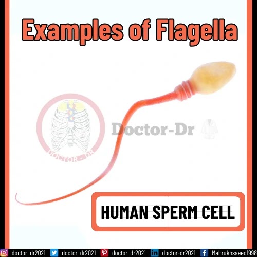
2. Flagellum in Human Sperm Cell:
- The flagellum in a human sperm cell is indispensable for motility and plays a vital role in in vivo fertilization.
- Successful propulsion of the cell, facilitated by the flagellum, is critical for ensuring successful fertilization during sexual reproduction in humans.
- Comprising microtubules arranged in a 9+2 pattern, the core of the flagellum includes structures such as the dynein regulatory complex, radial spokes, and dynein arms.
- The flagellum is essential for the penetration of the sperm into the egg during fertilization, providing the necessary orientation for the sperm in a specific direction.


