Table of Contents
- Introduction of Electron Microscope
- Working Principle of Electron Microscope
- Types of Electron microscope
- Parts of Electron Microscope
- Applications of Electron microscope
- Advantages of Electron microscope
- Limitations of Electron microscope
Introduction of Electron Microscope
An electron microscope is a specialized microscope that employs a beam of accelerated electrons as its light source. It offers extremely high image resolution, capable of magnifying objects to the nanometer scale by manipulating electrons in a vacuum and displaying the images on a phosphorescent screen.
The first electron microscope was developed in 1931 by German engineer and academic Ernst Ruska (1906-1988). The foundational principles of his design continue to shape modern electron microscopes today.
Working Principle of Electron Microscope
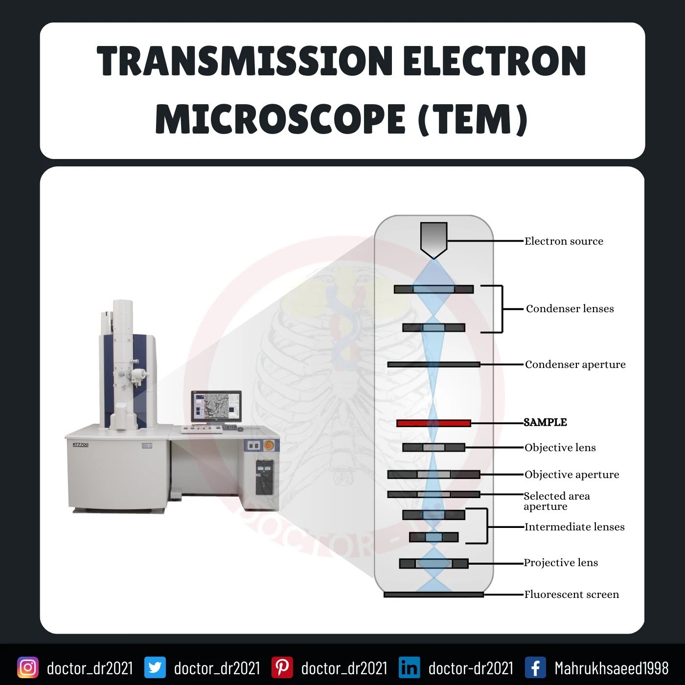
Electron microscopes gather data on the structure, morphology, and composition of a sample by analyzing signals generated from the interaction between the electron beam and the specimen.- The electron gun emits a stream of electrons.
- Two sets of condenser lenses focus this electron beam onto the specimen, forming a narrow and precise beam.
- To propel electrons down the column, an accelerating voltage (typically ranging from 100 kV to 1000 kV) is applied between the tungsten filament and the anode.
- The specimen being examined must be extremely thin, at least 200 times thinner than those used in optical microscopes. Ultra-thin sections, typically 20-100 nm thick, are placed on the specimen holder.
- As the electron beam passes through the specimen, electrons scatter based on the thickness or refractive index of different specimen regions.
- Denser areas in the specimen scatter more electrons, appearing darker in the final image since fewer electrons reach that part of the screen. In contrast, less dense, transparent regions appear brighter.
- The electron beam that exits the specimen is directed through the objective lens, which has high magnifying power and produces an intermediate magnified image.
- Finally, the ocular lenses further magnify the image to produce the final visual output.
Types of Electron microscope
1. Transmission Electron Microscope (TEM)
- The transmission electron microscope is designed to observe thin specimens that allow electrons to pass through, creating a projection image.
- In many ways, TEM operates similarly to a conventional compound light microscope.
- TEM is commonly used to visualize the internal structure of cells (in thin sections), the arrangement of protein molecules (enhanced by metal shadowing), the organization of molecules in viruses and cytoskeletal filaments (using the negative staining technique), and the structure of protein molecules in cell membranes (via freeze-fracture methods).
2. Scanning Electron Microscope (SEM)
- Conventional SEM relies on the emission of secondary electrons from the surface of a specimen.
- With its large depth of focus, SEM is the electron microscopy equivalent of a stereo light microscope.
- It provides highly detailed images of cell surfaces and entire organisms, which are not achievable through TEM. Additionally, SEM is useful for particle counting, size determination, and process control.
- It is called a scanning electron microscope because the image is generated by scanning a focused electron beam across the specimen's surface in a raster pattern.
Parts of Electron Microscope
The electron microscope consists of a vertically mounted tall vacuum column with the following key components:
1. Electron gun
- The electron gun is a heated tungsten filament responsible for generating the electron beam.
2. Electromagnetic lenses
- The condenser lens focuses the electron beam onto the specimen, while a second condenser lens narrows it into a fine, tight beam.
- After passing through the specimen, the electron beam moves through the objective lens, a high-power magnetic coil, which produces an intermediate magnified image.
- The third set of magnetic lenses, known as projector (ocular) lenses, further magnify the image.
- Each of these lenses enhances the image magnification while preserving an exceptional level of detail and resolution.
3. Specimen Holder
- The specimen holder consists of a thin film, typically made of carbon or collodion, supported by a metal grid.
4. Image viewing and Recording System
- The final image is displayed on a fluorescent screen, with a camera positioned below the screen to capture and record the image.
Applications of Electron microscope
- Electron microscopes are widely used to examine the ultrastructure of various biological and inorganic specimens, including microorganisms, cells, large molecules, biopsy samples, metals, and crystals.
- In industry, they are commonly employed for quality control and failure analysis.
- Modern electron microscopes utilize advanced digital cameras and frame grabbers to produce detailed electron micrographs.
- The field of microbiology owes much of its progress to electron microscopy, as it has significantly advanced the study of microorganisms such as bacteria, viruses, and other pathogens, greatly improving disease treatment.
Advantages of Electron microscope
- Extremely high magnification
- Exceptionally high resolution
- Minimal distortion of material during preparation
- Ability to explore a greater depth of field
- Versatile applications across various fields
Limitations of Electron microscope
- Live specimens cannot be observed.
- Due to the low penetration power of the electron beam, the specimen must be ultra-thin, requiring drying and slicing into very thin sections before examination.
- Since the electron microscope operates in a vacuum, specimens must be completely dried.
- It is costly to construct and maintain.
- Requires specialized training for researchers to operate.
- Image artifacts may occur due to specimen preparation.
- The microscope is large, bulky, and highly sensitive to vibrations and external magnetic fields.


